Basic Heart Anatomy and Physiology: Introduction to the Heart Chambers
Dive into the cardiac field with this introductory article about the generalities of the heart chambers, their functions, and their roles in the heart. Sparking a foundation for further readings about more in-depth structures about the heart.
ANATOMY AND PHYSIOLOGY
Avya Patel
3/8/20252 min read
Introduction to the Basics of the Heart
Think about the heart as a small pump, pushing blood through your body, and keeping your organs functioning. Pumping around 1.5 gallons of blood every minute, the heart is an incredibly complex organ with a very tedious task, if the heart does not function as it is supposed to, no other organ can take over its role. Now imagine this, the heart beats around 80 times a minute, 100,000 times a day, 35 million times a year, and around 2.5 billion times in an average lifespan, it is under constant stress, and because of this, it is important to understand the basic structure of the heart and how it works.
What does the heart look like and where is it located?
The heart looks like an acorn. It is located slightly to the left of the breastbone between the 1st and 5th intercostal spaces (the spaces in between your ribs). The heart is around the size of your fist and weighs 10-12 ounces in adults.
How is the heart divided?
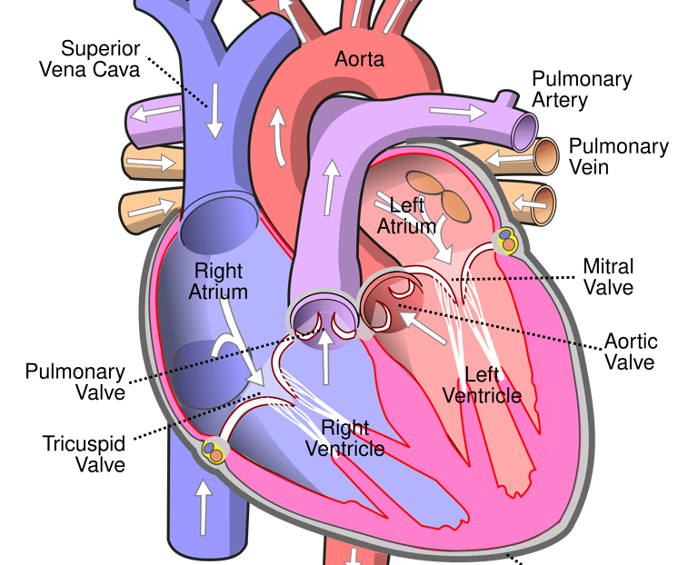
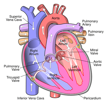
The heart is divided into 4 chambers, each with their respective roles.
The top right chamber is called the Right Atrium, abbreviated as ‘RA’.
The top left chamber is called the Left Atrium, abbreviated as ‘LA’.
The bottom right chamber is called the Right Ventricle, abbreviated as ‘RV’.
The bottom left chamber is called the Left Ventricle, abbreviated as ‘LV'.
A good rule of thumb is the Atrium/ Atria collects blood, while the Ventricle/ Ventricles send out blood. Arteries are blood vessels that send out blood from the heart, while veins are blood vessels that bring blood to the heart.
General details and roles of the chambers:
The right atrium (RA) collects deoxygenated blood from the two major veins called the Superior and Inferior Vena Cava. Blood flows from the Right Atrium to the Right Ventricle through the Tricuspid Valve (Tricuspid stems from Tri-three and Cuspid-tip/point, relating to 3 fibers part of the Tricuspid valve).
The right ventricle pushes deoxygenated blood it receives from the right atrium out to the lungs through the pulmonary valve via the pulmonary arteries, deoxygenated blood will then be oxygenated through small blood vessels called capillaries in the lungs. Here, Oxygen (O2) will replace Carbon Dioxide (CO2) in the blood cells, after this oxygenation phase, blood will be sent back to the left atrium through the pulmonary veins. Pulmonary arteries are the only arteries in the body that contain deoxygenated blood, while pulmonary veins are the only veins in the body that contain oxygenated blood.
The main role of the left atrium (LA) is to collect oxygenated blood from the lungs through pulmonary veins (*this is not shown in the image since pulmonary veins enter through the back of the left atrium) and then send it to the left ventricle (LV) through the opening of the mitral valve, also known as the bicuspid valve (Bicuspid stems from Bi-two and Cuspid-tip/point, relating to 2 fibers part of the Tricuspid valve).
Now finally, after collecting oxygenated blood from the left atrium, it is time for the left ventricle to send it out to the rest of the body. The left ventricle is responsible for delivering oxygenated blood to the rest of the body, once the aortic valve opens (connecting the left ventricle to the aorta), it sends blood from the left ventricle through the aortic branch and eventually, through the rest of the body. Once the blood is oxygenated blood used up by the body, it returns to the right atrium from the inferior and superior vena cava, and the whole process starts all over again.